Week 4: MedTech + Art
The most obvious
evidence of art in the medical world comes in the form of anatomy, where
artists and medical professionals have worked rigorously on correctly
identifying the internal structure and makeup within different species.
This overall structure includes muscles, bones, organs, and ligaments.
While doctors have performed the experiments required to correctly
identify the overall structure of bodies, artists are required to actually
realistically and accurately represent what doctors have proven from
experiment. One example of such artistic reconstruction can be seen at
the Bodies Exhibit, which displays 13 whole body human specimens at the Luxor
in Las Vegas (Luxor.com).
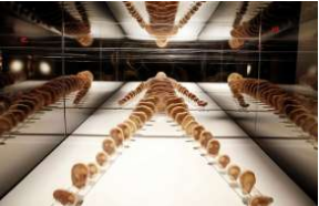 |
| Bodies Exhibit |
The advent of
computational technological advancement has allowed for further reconstruction
and imaging of internal body structure. Some examples of such imaging
includes X-Rays, MRIs, and CAT scans. I, personally, have undergone
dozens of X-Rays and MRIs during my lifetime, and have been fortunate enough to
look at and obtain copies of many of the images. X-Rays work by utilizing
electromagnetic waves to detect the bone structure of a species, with bones
showing up as white and other bodily organs appearing black (MedlinePlus.gov).
MRI scans work similarly by using magnetic and radio waves to detect
internal organs and tissues and create computerized images that display these
organs/tissues (Tidy).
 |
| X-Ray |
Recently, art and
medicine have further evolved to include plastic surgery and the insertion of
computer chips within the human body. One such example of the concept of
plastic surgery as art came up in this week’s lecture, and that is the example
of ORLAN. ORLAN is a French artist that uses herself as the artistic
medium. She has redefined her facial structure so that it aligns with
popular figures from historical art. For example, her forehead is
designed after da Vinci’s Mona Lisa
and her chin is designed to depict Botticelli’s Venus (ORLAN.edu).
 |
| ORLAN |
Kevin Warwick has been a
pioneer in the field of neuro-surgical implantations, as he has inserted an
implant into his left arm in order to link his nervous system to a computer in
an attempt to assist the disabled (Warwick). While experiments into this
field are fairly recent, they do give an idea of how art and technology can
help shape the field of medicine in the years to come; as well as an idea of
how far art and medicine have come since the early days of anatomy, to X-Rays
and plastic surgery, and finally to computerized implants.
References:
“Biography.” ORLAN, 2017,
www.orlan.eu/. Accessed 27 Apr. 2017.
“Bodies... The Exhibition.” Luxor,
2017,
www.luxor.com/en/entertainment/bodies-the-exhibition.html.
Accessed 27 Apr. 2017.
Tidy, Colin. “MRI Scan.” Patient, 23 Oct. 2015,
patient.info/health/mri-scan. Accessed 27 Apr. 2017.
Warwick, Kevin. “Kevin Warwick.” Kevin Warwick,
Coventry University, 2017, www.kevinwarwick.org/. Accessed 27 Apr. 2017.
“X-Rays.” MedlinePlus, U.S. National Library of
Medicine, 4 Mar. 2016, medlineplus.gov/xrays.html. Accessed 27 Apr. 2017.


Comments
Post a Comment